Some blood cancers are hard to tell apart, even though they may have significant differences.
Take the two most aggressive forms of leukemia: Acute myeloid leukemia (AML) and acute lymphocytic leukemia (ALL). Both start in bone marrow and are caused by an overabundance of abnormal, immature blood cells. They produce common symptoms in patients and have similar treatment options.
But AML and ALL are hardly the same and the prognoses for patients of the two diseases are vastly different.
AML has a 31.7 percent relative five-year survival rate. That’s the percentage of newly diagnosed AML patients expected to be alive five years later, compared to the percent of people without the disease who will still be alive over the same time period. ALL has a much higher survival rate of 71.3 percent, according to figures from the National Cancer Institute’s SEER database (Surveillance, Epidemiology, and End Results Program).
SEER data indicate that last year in the United States, there were 20,380 new cases of AML, which also goes by the names acute myeloblastic leukemia, acute myelogenous leukemia and acute nonlymphocytic leukemia. It caused an estimated 11,310 deaths in 2023.
The SEER numbers for ALL, also known as acute lymphoblastic leukemia, are lower, with 6,540 new cases and 1,390 deaths estimated in the United States last year.
In this article we will look at:
- Acute lymphocytic leukemia vs. acute myeloid leukemia
- Symptoms of ALL compared to AML
- AML and ALL causes and risk factors
- The ALL and AML diagnosis processes
- ALL vs. AML treatment differences
- Prognosis for AML compared to ALL
If you’ve been diagnosed with leukemia and have questions about your treatment plan, or you’re interested in a second opinion on your leukemia diagnosis, call us or chat online with a member of our team.
Acute lymphocytic leukemia vs. acute myeloid leukemia
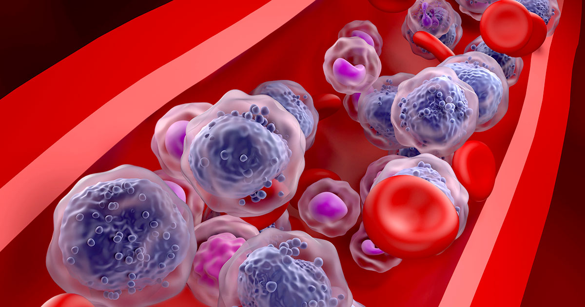
The human blood system is comprised of a variety of cells—red blood cells, white blood cells, platelets—and precursor cells in bone marrow that eventually develop into other cells.
Blood cancers, like other cancers, develop when something goes wrong with normal cell growth. For AML and ALL, the body produces too many immature precursor cells that impedes the development of normal blood cells and platelets.
For AML, these out-of-control immature cells, or blasts, are myeloid precursor cells. They would normally become a variety of cell types, including red blood cells, some specific versions of white blood cells—neutrophils, basophils, or eosinophils that engulf and dissolve bacteria and viruses—and platelets.
ALL involves lymphoid precursor cells, which predominantly develop under normal circumstances into two types of white blood cells: B-cell lymphocytes, which produce antibodies, and T-cell lymphocytes, which attack tumors and regulate immune system responses to infections.
“Both are very serious, life-threatening diseases that can affect people of all ages, although children are more likely to have ALL than AML,” says Leslie Popplewell, MD, Medical Director of HEM/BMT/Cellular Therapy in the Department of Hematology & Hematopoietic Cell Transplantation at City of Hope® Cancer Center Atlanta. “Both are treated with multi-agent chemotherapy, and often with allogeneic stem cell transplantation, which is from a family member or unrelated donor.”
Two related blood cancers start in more developed blood cells, which result in slower growing cancers. These are chronic myeloid leukemia (CML) and chronic lymphocytic leukemia (CLL).
Symptoms of ALL compared to AML
ALL and AML have mostly the same potential symptoms. Among the most common in both are:
- Fatigue
- Fever
- Night sweats
- Loss of appetite
- Feeling dizzy or light-headed
- Back or abdominal pain
- Pale skin
- Shortness of breath
- Tiny red dots under the skin
Some other symptoms are also possible in both, but they may be more linked to one disease than the other.
Those more likely to be AML symptoms are:
- Easy bruising or bleeding, including gum bleeding and nosebleeds
- Skin infections
- Bone pain
- Fullness in the stomach due to spleen or liver enlargement
Those typically more attributable to ALL are:
- Swollen lymph nodes in the neck, armpits, or groin
- Joint pain
- Nausea and vomiting
- Weight loss
- Enlargement of both the liver and spleen (hepatosplenomegaly)
ALL is also associated with headaches and the swelling of the thymus.
AML and ALL causes and risk factors
Many AML and ALL risk factors are similar. They include those listed below.
Prior chemotherapy: Previous cancer treatment with chemotherapy drugs is a risk factor for AML and ALL.
Radiation: Being exposed to high doses of radiation is a risk factor for both, as is exposure from previous radiation cancer treatments (although the latter risk is greater for ALL than AML).
Gender: Both cancers are more common among males, though male risk is greater with AML than ALL.
Environmental factors: Exposure to chemicals such as benzene may increase risk of both cancers, though the risk is greater for AML than ALL.
Genetic disorders: Some genetic disorders increase the risk for developing either AML or ALL, including Down syndrome, Fanconi anemia, Bloom syndrome and Ataxia-telangiectasia.
Other risk factors for developing the two blood cancers are more distinguishable. These risk factors include those listed below.
Age: AML is most likely to affect adults over the age of 65, while ALL is most common in children under the age of 15 and adults over 50.
Smoking: Exposure to smoking increases the risk for AML.
Family history of AML: Having an immediate family member with AML raises your risk for developing the disease.
Blood disorders: Having a history of a blood disorder—such as aplastic anemia, myelodysplastic syndrome or myeloproliferative neoplasm—is a risk factor for developing AML.
Race: White people are slightly more likely than Black people to develop ALL.
Viruses: ALL may be linked to a previous viral infection such as Epstein-Barr virus or human T-cell leukemia virus.
The ALL and AML diagnosis processes
Many of the same tools are used to diagnose AML and ALL, although sometimes for slightly different purposes. Here are some of the types of tests performed when these blood cancers are suspected or identified.
Blood tests
A series of blood tests may be ordered, including a CBC (complete blood count) that measures the number and types of cells in the blood. A blood smear observed under a microscope is also used to detect cell abnormalities. These will provide the first indications if leukemia is present.
Bone marrow tests
Patients may undergo two types of bone marrow procedures, which may be performed at the same time to assess the solid and liquid parts of the marrow. The procedures are a bone marrow aspiration, which uses a needle to remove a sample of the liquid marrow, and a bone marrow biopsy, which uses a needle to remove a sample of solid marrow tissue.
The samples are often taken from the pelvic bone under anesthesia, though the sternum (breastbone) or other bones may be used.
Cytochemistry and cytogenetic testing
After blood and tissue samples are collected from the patient, they may undergo cytochemistry (cytometry) examination to study cell size, cell DNA structure, cell morphology, cell proteins and the number of cells. Cytogenetic testing focused on chromosomes looks to see if there are broken ones, rearranged ones, missing ones or additional ones.
Spinal tap (lumber puncture)
A spinal tap, in which doctors remove some cerebrospinal fluid (CSF) from near the spinal cord, is performed to help determine if the leukemia has spread to the central nervous system (CNS). While used frequently for ALL patients, this is not a common AML test.
“There is more risk for patients with ALL to have involvement of the brain and/or cerebrospinal fluid, which can be difficult to treat,” Dr. Popplewell says. “Therefore, we are very proactive to give CNS-directed therapy to either treat CNS involvement or to prevent CNS involvement from the beginning.”
Imaging tests
Doctors may also order X-rays, CT scans or MRIs when investigating leukemia. Imaging scans of the pelvis and abdomen may help determine the stage of ALL. With AML patients, the imaging tests are typically used to spot infection, and not cancer spread, since AML is frequently not diagnosed until it is already at an advanced stage.
ALL vs AML treatment differences
The goal of treatment is to get rid of cancer cells, replenish normal blood cells and guard against the leukemia returning. The treatments for AML and ALL are similar, though not identical.
Leukemia treatments are often described in the following three phases.
Induction therapy: The first treatment after diagnosis that seeks to kill the cancer cells and put the patient into remission
Consolidation therapy: This treatment is a follow-up to the induction therapy, going after any remaining cancer cells
Maintenance therapy: Any treatment that may be necessary to control the cancer over an extended time period or keep the patient cancer free
“Between the two, ALL is often treated with a prolonged regimen, which can include induction, consolidation and then a prolonged maintenance phase which may go on for a few years,” Dr. Popplewell says. “AML is not treated with maintenance therapy generally once remission is achieved.”
Different treatments may be used in different phases, depending on the cancer and the success of any previous treatment. The treatments include those listed below.
Chemotherapy. This is typically the first line of defense against AML or ALL, seeking to put cancer into remission. The regimen may be more intense in younger patients who have a higher tolerance for chemotherapy.
Stem cell transplant. If the chemo doesn’t work, a patient may undergo a stem cell transplant (hematopoietic stem cell transplantation, or HSCT) that replaces the patient’s diseased bone marrow with healthy stem cells. Stem cell transplants are more common with AML than ALL, which typically relies more on multidrug chemotherapy after the initial chemotherapy.
Targeted therapy. These treatments use drugs that are designed to target and block specific cell abnormalities, with the goal of causing the cancer cells to die. AML and ALL generally use different forms of targeted therapies, based on different abnormalities.
Radiation therapy. This therapy, which uses high-energy beams such as X-rays to kill cancer cells, may be used as part of treatment. For AML patients, the radiation may be used to attack cancer cells that have spread to other parts of the body or in preparation for a stem cell transplant.
Radiation for ALL is sometimes directed toward the brain because of ALL’s potential to spread there from cancerous cells around the spinal column.
Prognosis for AML compared to ALL
AML has a much lower five-year survivability rate than ALL.
But survival rates vary even within a disease, based on factors like a person’s age, general health, biological attributes and how well he or she responds to treatment. ALL’s 71.3 percent relative five-year survival rate drops to 43 percent for people age 20 or older; it rises to 90 percent for those under age 20, according to information from the American Society of Clinical Oncology (ASCO).
AML’s 31.7 percent relative five-year survival rate drops to 28 percent for those age 20 and older, but it increases to 69 percent for those under 20.
If you’ve been diagnosed with leukemia and have questions about your treatment plan, or you’re interested in a second opinion on your leukemia diagnosis, call us or chat online with a member of our team.