This page was reviewed under our medical and editorial policy by
Henry Krebs, MD, Interventional and Diagnostic Radiologist
This page was reviewed on December 2, 2022.
If you have a growth that may be cancerous, your doctor may recommend getting a tissue sample to confirm whether it’s malignant and to determine how to treat it. A core needle biopsy allows doctors to do this without resorting to surgery. It’s often used to diagnose breast cancer.
A core needle biopsy obtains a cylinder of tissue about 1/16th of an inch in diameter. The needle is wider than that used for fine-needle aspiration (another form of needle biopsy), but since a core needle biopsy gathers more material, it means more information is derived.
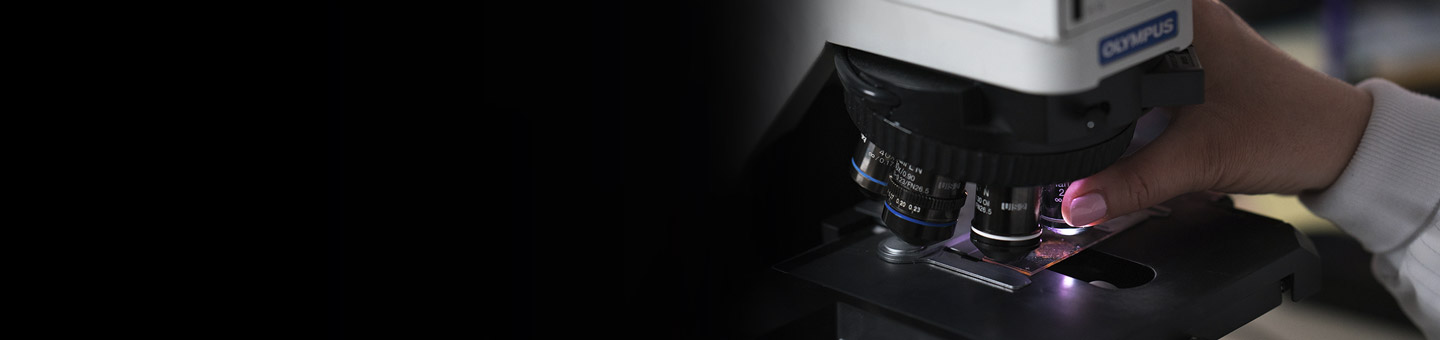
Fine-needle aspiration (FNA) samples undergo cytological examination, which looks at single cells or small groups of cells, while core needle biopsies allow a pathologist to perform a histological study of tissue structure and cells.
For breast cancer, a core needle biopsy may determine a tumor’s type and whether it’s likely to grow slowly or quickly, as well as whether it may respond to hormone therapy.
A core needle biopsy may be performed in your doctor’s office. In general:
Doctors often take several cores (samples) to get enough material. A doctor may also place a metal marker, called a clip, in the mass, so it may be easily located if surgery is needed. The clip is typically removed during surgery. If the patient doesn’t require surgery, the clip may be left inside (it’s considered harmless).
Once the procedure ends, pressure may be applied to limit any bleeding, and a bandage is placed over the insertion site. Doctors use this basic method of core needle biopsy for masses they are able to easily feel through the skin.
For growths or suspicious areas that can’t be felt, or palpated, through the skin, a doctor may use ultrasound to guide the needle to the growth. Sometimes a special vacuum helps extract the sample through the needle.
Procedures that use a needle or probe attached to a vacuum suction device are known as vacuum-assisted breast biopsies. While originally considered a different procedure from a core needle biopsy, the two techniques have become so similar that little distinction remains. The use of a vacuum may help collect additional tissue.
For some breast mass biopsies, more elaborate imaging techniques may be used, such as magnetic resonance imaging (MRI) or stereotactic mammography. Both may require that you lie facedown on a special table, with the procedures performed through a hole underneath. And both typically take place in a hospital radiology department.
There’s little preparation for most core needle biopsies. As with other medical procedures, tell your doctor in advance about:
Your doctor may advise you not to take aspirin, herbal preparations and blood-thinning medications such as Coumadin® (warfarin sodium) a few days before the appointment.
Ask your doctor whether the procedure will include sedation, in which case you’ll need a ride home.
Choose loose clothing (without metal fasteners if you’re having an MRI-guided procedure) and avoid wearing jewelry on the day of your appointment.
After the procedure:
Biopsy results are:
If positive, the pathology report may describe the type of cancer, the tumor grade (how abnormal the cells appear), and whether it’s contained or infiltrating neighboring tissue. Generally, the more abnormal and disorganized the cancer cells, the more likely they are to rapidly grow and spread. The sample also may be tested for sensitivity to hormones and other chemical characteristics, which helps paint a fuller picture of the cancer and the appropriate treatments.
A core needle biopsy provides more material, and therefore more information, than an FNA—but less material than a surgical biopsy. For breast lesions, a core needle biopsy may be performed quickly, with a low risk of infection or excessive bruising.
Samples may take longer to process than those from an FNA.
It’s also possible for the core needle to miss the suspected tumor, creating a false negative result. In image-guided core needle biopsies where the mass isn’t palpable (able to be felt), false negatives may happen in up to 4 percent of cases, according to Susan G. Komen. The rate is lower for palpable growths.
Other cons include:
Because a core needle biopsy collects a column of tissue, your care team may be able to tell whether the cancer is invasive or not—and, if used on lymph nodes, it may help in diagnosing some lymphomas. Diagnoses from core needle biopsies are more accurate and specific than those from an FNA, and your doctor may choose this technique if FNA results were inconclusive or incomplete. However, if a core needle biopsy doesn’t provide all the information your doctor needs, a surgical biopsy may be ordered.
Although core needle represents the preferred method to biopsy most breast masses, an FNA may be used for patients with cystic lesions or with a low likelihood of cancer.
In addition to breasts and lymph nodes, a core needle biopsy may be used to sample lung, kidney and other organ tissue, and sometimes musculoskeletal soft-tissue lesions.