This page was reviewed under our medical and editorial policy by
Daniel Liu, MD, Plastic and Reconstructive Surgeon
This page was reviewed on February 18, 2022.
You learned the stage of your breast cancer, but do you understand what it means?
Cancer stages involve myriad measurements and factors, making the process tricky to unravel—but vital.
Understanding your stage will help you and your care team make informed treatment decisions, including whether a clinical trial might be best suited for you.
Medical advances have broadened our knowledge about breast cancer and how to stage it, and those revelations have yielded improved treatments, too.
This article will cover:
Your breast cancer stage indicates the severity of the disease upon diagnosis. Your cancer stage will always stay the same, even if the cancer shrinks or spreads during or after treatment. For instance, if you’re diagnosed with stage 1 breast cancer, but the tumor later grows and spreads, it’s not considered stage 3 or 4 breast cancer. To determine whether the cancer has responded to treatment, a new stage may later be assigned an “r” in front of it to show that it’s different from the original stage.
Breast cancer staging is classified by:
All these attributes help your care team determine how to treat your cancer.
To assess the location, size and spread of cancer, your care team will use the TNM Staging System, developed and updated for breast cancer by the American Joint Committee on Cancer (AJCC).
Breast cancer has five general stages under the TNM system: 0 through 4
Stage 0 breast cancer, known as carcinoma in situ, means cancer cells are present but have been caught so early that they haven’t spread. In the ducts, it’s called ductal carcinoma in situ (DCIS) and referred to as noninvasive.
Stage 1 breast cancer means the tumor is very small and either has not spread or may have a tiny bit of spread in a nearby lymph node. A cancer that has spread into the surrounding area is referred to as invasive breast cancer.
Stage 2 breast cancer means the tumor is larger than at stage 1 and may have spread to a few nearby lymph nodes.
Stage 3 breast cancer means the tumor is larger than at stage 2 and/or has spread to several lymph nodes and/or tissue around the breast or breast bone.
Stage 4 breast cancer means the cancer has metastasized, or become mobile, and spread to distant parts of the body, typically the bones, lungs or liver. This is an advanced stage of cancer, called metastatic breast cancer.
Recurrent breast cancer occurs when the disease has returned after initial treatment. Most recurrent cancers appear within the first two or three years after treatment, but, in some cases, the cancer may recur many years later.
Your cancer will be clinical staged based on diagnostic tests, a pathological staged after surgery (which allows your care team to closely study the cancerous tissue).
TNM staging involves quite a bit of detailed information. Let’s break down this system:
T refers to the size of the tumor.
An imaging technique may be used to measure the tumor. The designations are:
N refers to spread to nearby lymph nodes.
Lymph nodes are pockets in your lymphatic system, which is similar to the circulatory system. Instead of blood, it transports a fluid called lymph. White blood cells are key immune cells that cluster in your lymph nodes, waiting to defend against foreign invaders. Cancer can spread to these nodes.
The cancer found in lymph nodes may be small (micrometastasis: 0.2 mm to 2 mm) or large (macrometastasis: bigger than 2 mm). The designations are:
M refers to the spread to more distant parts of the body, or metastasis.
Additional markers specific to breast cancer will further define your stage, which may be helpful in choosing targeted treatments to fight the cancer.
These markers, along with the TNM measurements, define your stage.
A cancer recurrence refers to cancer that returns in the same breast, and it requires new staging. This new stage is marked by an “R” at the end to indicate “restaging.” If it develops in the other breast, it’s considered a new cancer.
Your breast cancer stage is specific to you. It’s based on all the factors collected through diagnostic tests and/or surgery, and it helps your care team assess your prognosis.
The American Cancer Society offers examples of how to interpret a breast cancer stage. For example, a cancer that’s T2, grade 3, no spread to lymph nodes (N0) or body (M0), HER2-negative and ER- and PR-positive is stage 1B.
Many methods are used to detect and stage cancer. Some of the common tests include:
Biopsy: The doctor uses a needle to extract breast tissue or fluid, which is then sent to a lab. There, various techniques are used to examine different attributes, such as hormone receptor or HER2 status. (Sometimes the tissue sample is collected during a surgical procedure informing the pathological stage.)
Tumor markers: Rapidly dividing cancerous cells interrupt some of the normal mechanisms of cell growth. This causes the cell to overproduce certain molecules. Lab tests detect these compounds, known as tumor markers, in blood or tissue samples.
Imaging techniques: Several different scans are used to examine characteristics of your cancer. Below are some of the noninvasive imaging techniques you might encounter:
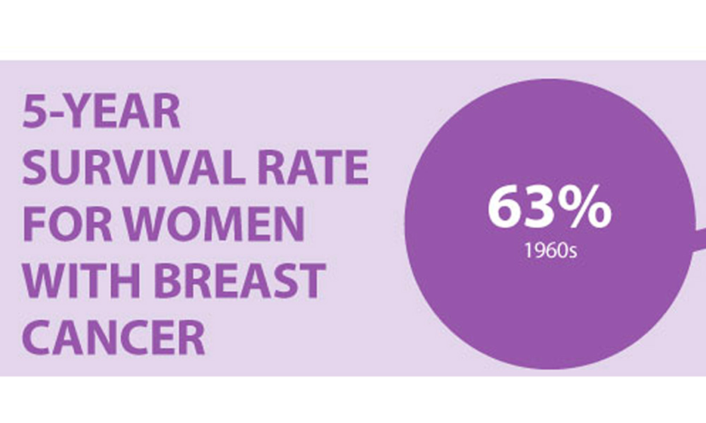
According to the National Cancer Institute (NCI), the percentage of patients surviving five years after diagnosis is:
The NCI also lists the five-year survival rate for breast cancer overall as 90.6 percent for women and 83 percent for men.
Download breast cancer infographic »
During your initial diagnosis, you and your cancer team will work together to develop a treatment plan. Staging allows you to answer the following questions:
Some of the staging may be even more in-depth, but in general, it’s designed to prepare a more tailored approach to your disease. Your care team will be able to explain any new terms and what they mean for you.